What Are Thyroid Nodules?
What are thyroid nodules? Basically, they are lumps in the thyroid. The thyroid is a butterfly shaped gland in the lower, anterior area of your neck. It secretes thyroid hormone that controls metabolism and protein production throughout the body. Many studies find that nodules are common. Luckily most do not become clinically significant, but about 5% can be a thyroid cancer.
Types of Nodules
Nodules can be benign (noncancerous) or malignant (cancerous). There are several types of benign nodules.
Benign nodules:
Hyperplastic nodules are made up of abnormal over growth of normal cells.
Colloid nodules are partly solid and partly cystic. The cystic areas are filled with a fluid called colloid, which is created when the thyroid produces thyroid hormone. A pure cyst can cause a lump and is rarely a cancer.
An adenoma is a benign tumor. Sometimes the difference between an adenoma and a cancer (carcinoma) can only be determined after surgical removal. Hashimoto’s thyroiditis, an autoimmune inflammation of the thyroid gland, can frequently cause a nodular goiter.
A multinodular goiter is a thyroid with multiple nodules, whereas a uninodular goiter is made up of only one nodule. The word “goiter” is a term that means enlarged thyroid. This can be caused by one or multiple nodules or by diffuse enlargement of the thyroid gland
Malignant nodules:
As noted above, about 5% of nodules can be malignant. There are several types of thyroid carcinoma (cancer).
Papillary thyroid carcinoma is by far the most common, making up about 80%.
Follicular thyroid carcinoma is the second most common. It can only be differentiated from a benign follicular adenoma by removal and examination of the entire tumor by a pathologist.
Anaplastic carcinoma makes up less than 5% of thyroid cancer. It is very fast growing and very aggressive. Treatment is difficult and the prognosis is poor.
Another uncommon cancer (< 5%) is called medullary thyroid carcinoma. It can be linked to genetic risk factors.
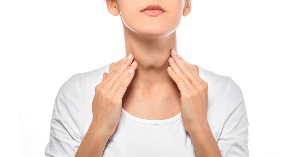
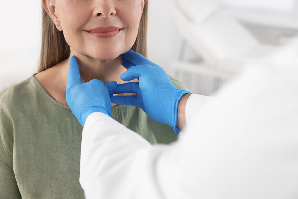
Nodules are often found by doctors during a physical exam. Some can be seen on the front of the neck and are brought to someone’s attention when someone notices it moving with swallowing or talking. Others are found coincidentally on imaging studies ordered to evaluate other problems, such as neck pain, lung problems, carotid artery blockage, etc.
Thyroid stimulating hormone (TSH)
The evaluation of the lesions in the thyroid is best done with ultrasound. Sometimes a CT (CAT) scan or magnetic resonance imaging (MRI) is used. A blood test to evaluate the thyroid stimulating hormone (TSH) level will tell if the thyroid is functioning normally.
If the TSH is low, meaning the thyroid is over functioning, then a radioactive iodine uptake and scan needs to be done. Once a nodule is located in your thyroid, a discussion with your doctor will determine the best course of action for you. The decision to do a biopsy of the nodule is determined by size and characteristics of the nodule seen on the ultrasound.
Symptoms of Thyroid Nodules
Typically, thyroid nodules do not present with any symptoms. They are found coincidentally on imaging or on physical exam in a provider’s exam. Sometimes a patient will find a nodule with feeling the neck or looking in the mirror. The presence of symptoms is an indication for surgery.
A nodule can be seen in the front of the lower neck. They can be seen in the mirror or noticed by other people. They can be more noticeable during talking or swallowing.
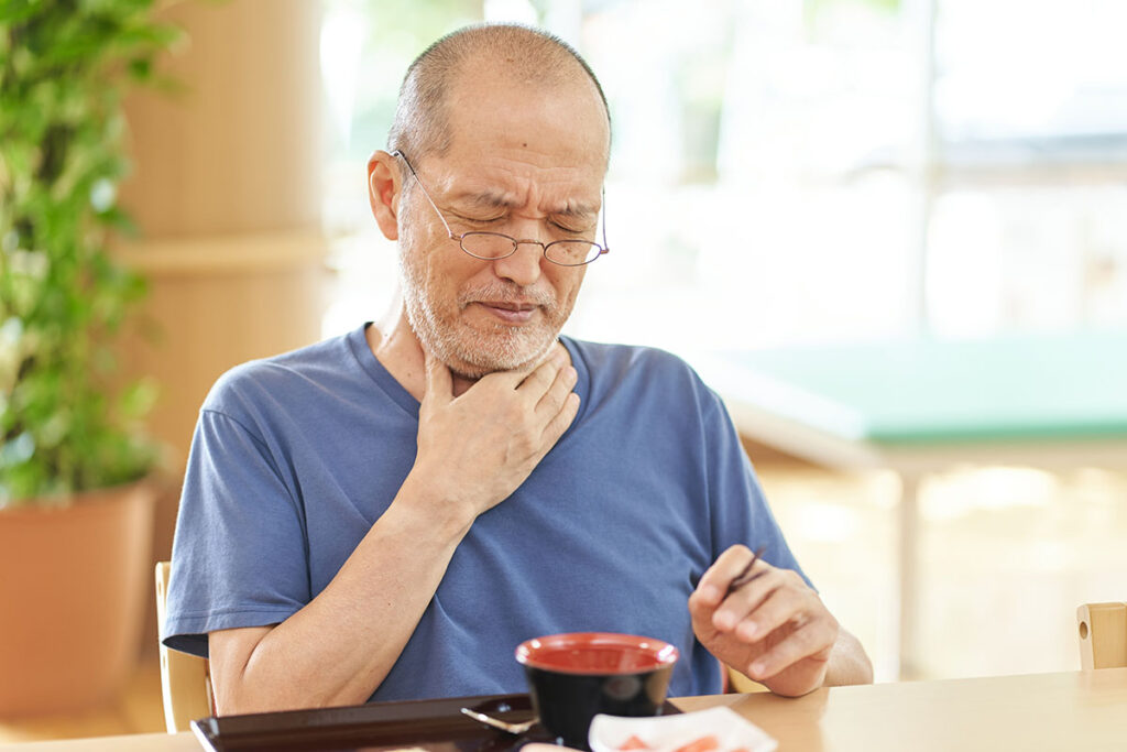
As nodules get larger there is a higher chance of symptoms as they push on surrounding structures. Pressure on the throat or upper esophagus can cause problems swallowing. Food feels like it catches in the lower neck. It feels like you have to swallow a couple times or sip water to help foods go down. Some nodules can be seen in the front of the neck. If the nodules are causing symptoms, or if they are visible they can be treated with surgery or Radio Frequency Ablation.
As nodules push on the trachea (windpipe) there can be tightness in breathing. This is typically not dangerous unless enlargement on both sides cause narrowing by pinching the trachea. Pressure on the nerve to the voice box can cause hoarseness. A complete loss of voice and weakness in the voice or cough can be caused by pressure on the nerve or by cancer affecting the nerve. This is uncommon.
Some people will feel tightness in the neck or pressure sensation when lying back or in certain neck positions.
Sometimes a nodule will secrete high levels of thyroid hormone, called a toxic nodule, causing hyperthyroidism. This is determined with a blood test to look at thyroid hormone levels. If the thyroid stimulating hormone (TSH) level is low, indicating high levels of thyroid hormone, then you will need a radioactive iodine uptake scan to help determine the cause of the high thyroid hormone levels. Some symptoms of hyperthyroidism are heat intolerance, high heart rate, tremors, weight loss, dry hair and skin, and anxiety.
Request an Appointment
Same and next day appointments available